Image Guided Medialization Laryngoplasty
Vocal cord paralysis and paresis are debilitating conditions leading to difficulty with voice production. Medialization laryngoplasty is a surgical procedure designed to restore the voice in patients by implanting a uniquely configured structural support lateral to the paretic vocal fold through a window cut in the thyroid cartilage of the larynx. Currently, the surgeon relies on experience and intuition to place the implant in the desired location, therefore it is subject to a significant level of uncertainty. Window placement errors of up to 5mm in the vertical dimension are common in patients admitted for revision surgery.

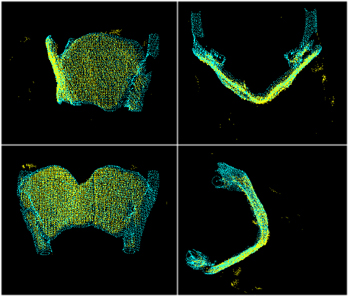
The failure rate of this procedure is as high as 24% even for experienced surgeons. An intraoperative image-guided system will help the surgeon to accurately place the implant by superimposing the CT data from the patient with the actual larynx of the patient during surgery. One of the fundamental challenges in our system is to accurately register the preoperative 3D CT data to the intraoperative 3D surfaces of the patient. Our proposed image guided system will use the anatomical and geometric landmarks and points to register intraoperative 3D surface of thyroid cartilage to the preoperative 3D radiological data. The proposed approach has three phases. First, the laryngeal cartilage surface is segmented out from the preoperative 3D CT data. Second, the surface of the exposed laryngeal cartilage during the surgery is reconstructed intraop-eratively using stereo vision and structured light based surface scanning. Third, the two geometries are registered using ICP based shape matching. The proposed approach has several advantages over alternative approaches: the combination of stereo vision and structured light surface scanning is capable of tracking the fiducial markers, reconstructing the surface of laryngeal cartilage and matching the preoperative and postoperative surfaces for registration purposes. The computer vision based approach can be applied to delicate areas like laryngeal cartilage with no danger of causing physical damage.
Participants: Steven Bielamowicz, M.D., James Hahn, Ph.D., Rajat Mittal, Ph.D., Raymond Walsh, Ph.D.

