
Vena Cava Filter Placement
CT or MR images acquired from the patient are used to automatically segment out the vasculature which is then stored in a hierarchical blood vessel data structure. This information is used by the simulation module to perform a dynamic simulation of a catheter moving through the vasculature.
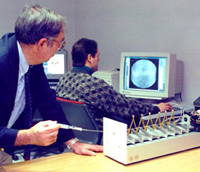
A proprietary force-feedback device has been developed by the mechanical engineering department. This device relays user movements to the simulation, which computes the catheter interaction with the vasculature, and returns the proper forces and torques to the device, which then outputs them to the user.
The rendering module accesses the blood vessel data structure, the catheter data structure, as well as an environment map to produce a fluoroscopic representation on a display screen. The entire system operates at interactive frame rates.
The catheter simulator has been used in the creation of a tutorial and training simulation of Inferior Vena Cava (IVC) filter placement. The filter is guided into place using a catheter inserted through an incision at a remote location. The procedure is monitored through a fluoroscopic view of the patient presented on a screen in front of the surgeon.
Participants: James K. Hahn, Roger Kaufman, Raymond Walsh, Adam Winick, Thurston Carleton, Nadia al-Ghreimil